Treatment of endometriosis

Endometriosis: A shared challenge

10% of women in reproductive age are affected by endometriosis. [1]

In many cases, surgical treatment becomes necessary. Depending on the extent and location of the disease, procedures can range from treating superficial lesions to operating in complex and critical anatomical areas.

Erbe offers devices and instruments for uniform tissue effects, adaptable hemostasis, and efficient dissection. Solutions such as TriSect rapide®, BiClamp®, and APCapplicator are designed to support surgical workflows and facilitate work in and around sensitive structures. Discover how these solutions enhance the surgical treatment of endometriosis.
 

Contact us

Endometriosis occurs in different phenotypes

Depending on the phenotype, endometriosis can involve structures that require careful dissection, reliable hemostasis, and controlled energy application.

Deep infiltrating endometriosis often involves dense adhesions and lesions close to critical structures. Effective excision requires precise energy delivery, reliable tissue control, and minimal lateral thermal spread.

Solutions that support consistent performance and dependable hemostasis are essential in these challenging conditions. TriSect rapide enables dissection close to critical structures, with a lateral thermal spread of less than 1 mm.

What experts say

The TriSect rapide® is an instrument that makes surgery easier when we treat deep endometriosis, particularly parametrial disease, which – along with many of my colleagues – I consider to be the most challenging form of endometriosis. Some surgeons argue that when deep endometriosis involves the lateral part of the parametrium, the vessels cannot be spared when removing that tissue completely. I disagree. With the right instrument, a thorough understanding of the anatomy, and the optimal technique in hand, the TriSect rapide® allows us to preserve those vessels. This is crucial for women who wish to retain their uterus for future pregnancy. Moreover, the device enables safe, controlled, yet highly effective hemostasis, as demonstrated in the videos that showcase its combined dissection and thermal capabilities.

Dr. Carlo Alboni
Dr. Carlo Alboni

Modena, Italy

No video found or Vimeo API error.

How Erbe supports your workflow

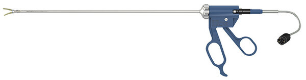
TriSect rapide®

TriSect rapide®

Dissect and seal in a true single step.

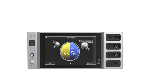
VIO® 3n Water

VIO® 3n Water

Designed for complex cases in multi-disciplinary ORs.

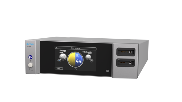
VIO® seal

VIO® seal

Generator entirely dedicated to bipolar applications.

BiClamp® LAP forceps

BiClamp® LAP forceps

Sealing of vessels and tissue structures for laparoscopic procedures.

Products in focus

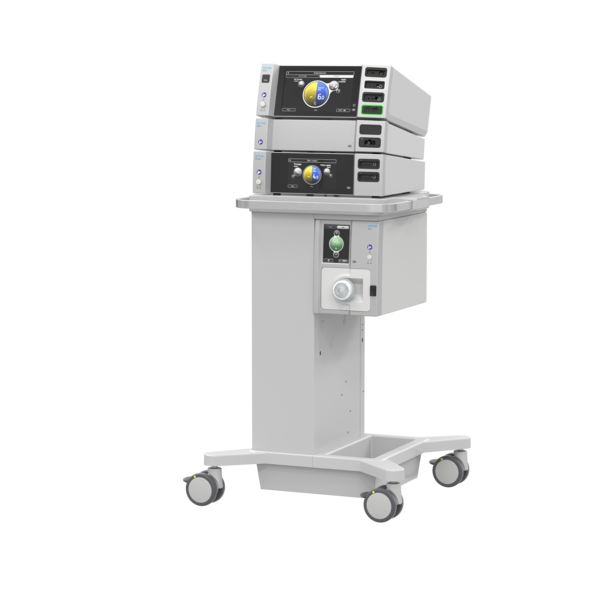
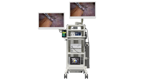
Workstation VIRON X System

Scientific information

Summary of publication

Muzii, Ludovico et al. “Histologic analysis of endometriomas: what the surgeon needs to know” Fertility and Sterility vol. 87.2 (2007):

Key findings

  • The inner cyst wall was lined by endometriotic tissue over a median of 60% (range 10-98%) of the surface.
  • The mean cyst wall thickness was 1.4 mm. Endometriotic tissue penetrated the cyst wall to a mean maximal depth of 0.6 mm (range: 0.1–2.0 mm). In 99% of cases, tissue penetration did not exceed 1.5 mm, and in 87% of cases, it did not exceed 1.0 mm.

Conclusion


The study demonstrates the extent and depth of endometriotic tissue spread in the endometrioma cyst.  Any treatment must consider the full spread of endometriotic tissue in the cyst wall, while preserving underlying healthy ovarian tissue.

Read more

Summary of publication

Stefanovic, Stefan et al. “Microscopic, Macroscopic and Thermal Impact of Argon Plasma, Diode Laser, and Electrocoagulation on Ovarian Tissue” In Vivo vol. 37 (2) 531-538 (2023):

Key findings

  • After 5 seconds, bipolar electrocoagulation caused overheating (>40°C) in 41.7% of samples.
  • Adjacent tissue heating was least pronounced with preciseAPC® and monopolar electrocoagulation. With preciseAPC® mode after 5 sec activation a mean of 27.2 ± 3.3°C was reached.
  • preciseAPC® produced the shallowest defects of all modalities, with a mean depth of only 0.05 mm after 5 seconds.

Conclusion
The study demonstrated favorable thermal tissue effects with preciseAPC®, including minimal tissue penetration and limited adjacent tissue heating in an ex vivo bovine ovarian model. These findings support further investigation of preciseAPC® for ovarian tissue-preserving surgical applications.

Summary of publication


Ferrari, Stefano M. et al. “Histologic findings from ablation of ovarian endometriomas using argon plasma coagulation (APC-ENDO): a pilot study” European Journal of Obstetrics & Gynecology and Reproductive Biology vol. 312 (2025):

Key findings

  • Histologic analysis showed a mean coagulation depth of 0.8 mm (95% confidence interval 0.42–1.17 mm). 
  • The maximum observed depth was 1.5 mm, remaining within the thickness of the fibrotic pseudocapsule.
  • No residual endometrial epithelium or stroma was detected in any coagulated area, indicating complete ablation of endometriotic tissue.
  • No thermal damage extended into the surrounding ovarian parenchyma, demonstrating strict confinement of necrosis to the pseudocapsule.

Conclusion

Argon plasma coagulation using the APC2 system produces controlled, shallow coagulation confined to the endometrioma pseudocapsule, achieving complete ablation of endometriotic tissue while sparing adjacent ovarian parenchyma. These findings support APC as a promising, tissue-preserving alternative to cystectomy. Additional clinical evidence from San Raffaele Hospital indicates thatpreciseAPC effect 9.0 creates a tissue effect that is:
Deep enough to remove endometriotic tissue from the cysts
Superficial enough to preserve the ovarian tissue
Clinical studies evaluating long term clinical effects on fertility, recurrence, and pregnancy rate will be conducted.
 

Summary of publication


E. Saridogan et al., "Recommendations for the surgical treatment of endometriosis-part 1: ovarian endometrioma," Gynecol Surg, vol. 14, no. 1, p. 27, 2017

Key findings

  • Cystectomy (stripping technique): Recommended as an effective method for complete removal of endometriotic tissue but is associated with unavoidable loss of ovarian tissue and potential reduction in ovarian reserve.
  • Ablation techniques (laser or argon plasma): Carbon dioxide (CO₂) laser and argon plasma are recommended for treatment of endometriotic tissue confined to the superficial cyst lining. These techniques may better preserve ovarian parenchyma compared with cystectomy when performed correctly.
  • Electrocoagulation: Widely used but associated with deeper and less predictable thermal spread. Short application times, low power settings, frequent cooling, and cautious use are strongly emphasized.

Conclusion

This consensus document provides detailed, technically focused recommendations for the surgical management of ovarian endometriomas, complementing existing evidence-based guidelines. It underscores the need to balance complete eradication of endometriotic tissue with maximal preservation of ovarian function, for which for example APC may be a feasible approach.
 

FAQ

Deep infiltrating endometriosis (DIE) often requires extensive dissection near critical anatomical structures. Surgical treatment aims to achieve complete excision of lesions while preserving healthy surrounding tissue whenever possible.

Erbe supports these procedures with advanced electrosurgical instruments such as TriSect rapide®. The instrument combines dissection, sealing, and coagulation functions in a single device, helping surgeons perform precise tissue dissection and hemostasis during complex laparoscopic procedures.

TriSect rapide® offers low lateral thermal spread and rapid jaw cooling, which may support work in anatomically challenging areas where precision is important. In addition, the softCOAG® mode provides immediate hemostasis when required during dissection.

Electrosurgery plays an important role in the surgical treatment of endometriosis by supporting tissue dissection, coagulation, hemostasis, and lesion removal during minimally invasive procedures.

Depending on the type and location of the disease, surgeons may use different electrosurgical instruments to excise deep infiltrating lesions, perform adhesiolysis, coagulate bleeding vessels, or ablate superficial endometriotic tissue. The choice of energy source can influence surgical precision, tissue effects, and the preservation of surrounding healthy structures.

For complex procedures such as deep infiltrating endometriosis (DIE), Erbe's TriSect rapide® combines dissection, sealing, and coagulation functions in a single instrument. In ovarian and superficial endometriosis, argon plasma coagulation technologies such as the APCapplicator can support controlled coagulation and tissue ablation while maintaining effective hemostasis.

Argon plasma coagulation (APC ) is an electrosurgical monopolar procedure for superficial hemostasis, devitalization and ablation using ionized argon gas.

Erbe's APCapplicator is designed to support contact-free coagulation and ablation with ergonomic handling and low smoke plume development. In endometriosis surgery, APC can be used to treat ovarian and superficial lesions by creating a homogeneous tissue effect with a controlled depth of penetration. Recent studies have demonstrated favorable tissue effects and limited thermal impact on the surrounding ovarian tissue. 

Lateral thermal spread refers to the transfer of heat from the target tissue to adjacent structures during the use of electrosurgical instruments.

In endometriosis surgery, lesions are often located close to critical anatomical structures. Excessive thermal spread may increase the risk of unintended tissue damage beyond the intended treatment area.

For this reason, surgeons often seek technologies that provide precise energy delivery and controlled tissue effects. TriSect rapide® offers a lateral thermal spread of less than 1 mm, supporting precise dissection and sealing when operating close to the sensitive anatomical structures encountered in deep infiltrating endometriosis procedures.

Recent innovations in endometriosis surgery focus on improving precision, minimizing thermal tissue damage, and supporting fertility-preserving approaches.

One area of development is multifunctional laparoscopic instrumentation. Instruments such as Erbe's TriSect rapide® combine dissection, vessel sealing, and coagulation in a single instrument, helping surgeons manage complex procedures more efficiently.

Another innovation is the growing use of advanced argon plasma coagulation instruments. Erbe's APCapplicator with preciseAPC® technology enables controlled superficial tissue effects with limited penetration depth. Histological and ex vivo studies have shown promising results regarding tissue preservation and controlled coagulation depth.

Endometriosis is commonly classified into three main phenotypes based on the location and depth of the lesions:

Superficial peritoneal endometriosis (SPE)
This form affects the peritoneum, the thin membrane lining the pelvic cavity. Lesions are located on the tissue surface and are generally considered the least invasive form of the disease.

Ovarian endometriosis (OE)
Ovarian endometriosis is characterized by endometriomas, often referred to as ovarian cysts. Surgical treatment aims to remove or ablate endometriotic tissue while preserving healthy ovarian tissue and ovarian reserve whenever possible.

Deep infiltrating endometriosis (DIE)
Deep infiltrating endometriosis is the most complex phenotype. Lesions penetrate more than 5 mm beneath the peritoneal surface and may involve structures such as the bowel, bladder, ureters, pelvic nerves, or uterosacral ligaments. Surgical treatment often requires advanced dissection techniques and multidisciplinary expertise.

Because each phenotype presents different surgical challenges, treatment strategies and energy modalities may vary. 

References

  1. World Health Organization. Endometriosis [Internet]. Geneva: WHO; 2023 Mar 24 [cited 2025 Sep 29]. 
 Available from: www.who.int/news-room/fact-sheets/detail/endometriosis 
  2. E. Saridogan et al., “Recommendations for the surgical treatment of endometriosis-part 1: ovarian endometrioma,” Gynecol Surg, vol. 14, no. 1, p. 27, 2017, doi: 10.1186/s10397-017-1029-x